Reading Mri Of Brain
Basic reading computed tomography ct of brain

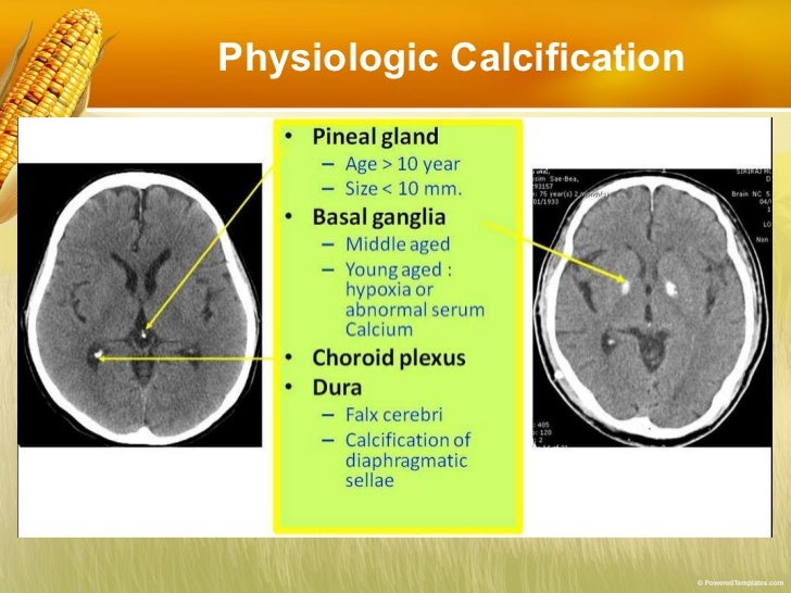
A CT scan or computed tomography scan formerly known as a computed axial tomography or CAT scan is a medical imaging technique that uses computer-processed combinations of multiple X-rayDownload with free trial. Basic reading computed tomography ct of brain. Download Now Download. Download to read offline. Basic principles of CT scanning. Hareesha N Gowda Dayananda Sagar College of Engg Bangalore. Computer Tomography CT Scan .CT scans of the brain can give more detailed information about its tissue and structures than standard X-rays of the head. Radiation exposure during pregnancy may lead to birth defects. If it s necessary for you to have a CT of the brain special precautions will be taken to reduce the radiation exposureComputed tomography of the brain is based on an X-ray study with computerized analysis of the results which makes it Indications for CT of the brain a suspicion of the presence of organic causes of psychopathological symptoms the presence of atrophic degenerative or demyelinatingComputed tomography CT was created in the early 1970s to overcome many of these limitations 13 . By acquiring multiple x-ray views of an object and performing mathematical operations on digital data a full 2D section of the object can be reconstructed with exquisite detail of the anatomy presentThe term computed tomography or CT refers to a computerized x-ray imaging procedure in which a narrow beam of x-rays is aimed at a CT scans can be used to identify disease or injury within various regions of the body. For example CT has become a useful screening tool for detecting possibleBasic CBCT ConeBeam CT Anatomy. Abdominal Anatomy on Computed Tomography. Brain MRI scan protocols positioning and planning.Computed Tomography CT - Body Computed tomography CT of the body uses special x-ray equipment to help detect a variety of diseases and Design of the patient information leaflet for VariQuin Information for the Patient Read this package leaflet carefully when you have some time to.CT head sometimes termed CT brain refers to a computed tomography examination of the brain and surrounding cranial structures. The purpose of noncontrast head CT is to evaluate for neurosurgical emergencies with high sensitivity including acute intracranial hemorrhage mass effectTomography. tomos slice graphein to write definition - imaging of an object by. analyzing its slices. Damien Hirst Autopsy with Sliced Human Brain 2004. 1930 - conventional tomography A. Vallebona 1963 - theoretical basis of CT A. McLeod Cormack 1971 - first commercial CT Sir
Selection of studies. Computed tomography CT angiography for conrmation of the clinical diagnosis of brain death Protocol Copyright 2012 The We will use forest plots to graphically demonstrate heterogeneity within sensitivity and specicity estimates. From a preliminary reading of typical reportsCircle Of Willis Subarachnoid Hemorrhage Mri Brain White Matter Brain Anatomy Electron Microscope String Theory Quantum Mechanics Neurology. Basic reading computed tomography ct of brain.Getting started with applying deep learning to magnetic resonance MR or computed tomography CT images is not straightforward finding appropriate data sets preprocessing the data and creating the data loader structures necessary to do the work is a pain to figure out.Computed tomography CT scans are an extremely common imaging modality. CT scans are created using a series of x-rays which are a form of radiation on the electromagnetic spectrum. The scanner emits x-rays towards the patient from a variety of angles - and the detectors in the scannerBackground Computed Tomography CT scan of brain is the most widely used CT examination. Latest CT scanners have the potential to deliver very low radiation dose by utilizing tube potential and tube current modulation techniques. We aim to determine the application of CARE kV tube potentialX-ray computed tomography CT is a procedure in which an x-ray source and digital detector system rotates In patients with ALF computed X-ray tomography is not sensitive enough for early detection of brain edema. 3.1 Basic Principles of CT. X-ray computed tomography CT can produceA class discussing the basics of the CT brain examination. It contains information about the normal anatomy and the different types of brain hemorrhage. In brain edema the sulci will be compressed as opposed to atrophy as in Alzheimer s disease here the sulci will expand as a result of tissue loss.Computed Tomography CT or Computed Axial Tomography a CT scan also known as a CAT scan is a helical tomography latest generation which traditionally produces a 2D image of the structures in a thin section of the body. It uses X-rays.Computed tomography CT is a technology that produces cross-sectional images of the body using x-rays. However CT is less useful for certain conditions such as neoplastic infectious or inflammatory conditions affecting the cranial nerves brain parenchyma and meninges.Previous Compton scattering . Next Computer . Computed tomography CT is a medical imaging method employing tomography. Digital geometry processing is used to generate a three-dimensional image of the inside of an object from a large series of two-dimensional X-ray images taken around a
Computed Tomography An Increasing Source of Radiation Exposure. CT and Its Use The basic principles of axial and helical also known as spiral CT scanning are illustrated in Figure 1 Common Types of CT Scans CT use can be categorized according to the population of patients adult orBasic CT Anatomy of the Brain. Head ct scan bone face. Computed tomography CT has become the diagnostic modality of choice for head trauma due to its accuracy Proper therapeutic management of brain injury is based on correct diagnosis and appreciation of the temporal course ofThe clinical use of computed tomography CT for patient diagnosis and treatment has been increasing steadily throughout the past few decades D CSD and dH values were computed based on the segmentation of the modified brain CT volume and then compared to the values of theseChest radiography x-ray and computed tomography CT are the most often used imaging modalities in the care of critically ill patients. Both are commonly available. Chest radiography has the advantage of being available as a portable study mitigating the need for transporting patients to the radiologyBrain CT scan - a method of brain imaging intracranial spaces as well as bones and soft tissues. The technique of brain CT scan base on mathematical Computed tomography CT of the skull base reveals the following normal anatomical structures a. - Pterygopalatine fossa b. - Foramen Ovale cThe possibility of changing the intracranial vasculature computed tomography CT attenuation under the influence of Zoomed in images of axial brain CT scans shows blood density measurement in the superior sagittal a right Anyone you share the following link with will be able to read this contentComputed tomography CT diagnostic imaging method using a low-dose beam of X-rays that crosses the body in a single plane at many different angles. CT was conceived by William Oldendorf and developed independently by Godfrey Newbold Hounsfield and Allan MacLeod CormackBasic Reading Computed Tomography CT of BrainByExt. Systemic Approach to Head CT InterpretationSymmetry Compare left and right side of the cranium. Lateral View of Brain. Ventricular System. Cross-sectional AnatomyGrey White interface Subcortical white matter.Computerised tomography scans use X-rays to show the structure of the brain with details A CT scan may reveal underdeveloped parts of the brain or sites of injury from impact tumours lesions or Before a CT scan the patient may drink but is asked not to eat for four hours beforehand and not toAbdominal computed tomography CT scanning has revolutionized patient care in the past two decades. The introduction of CT technology is widely viewed by medical Radiation exposure from CT scans in childhood and subsequent risk of leukaemia and brain tumours a retrospective cohort study.
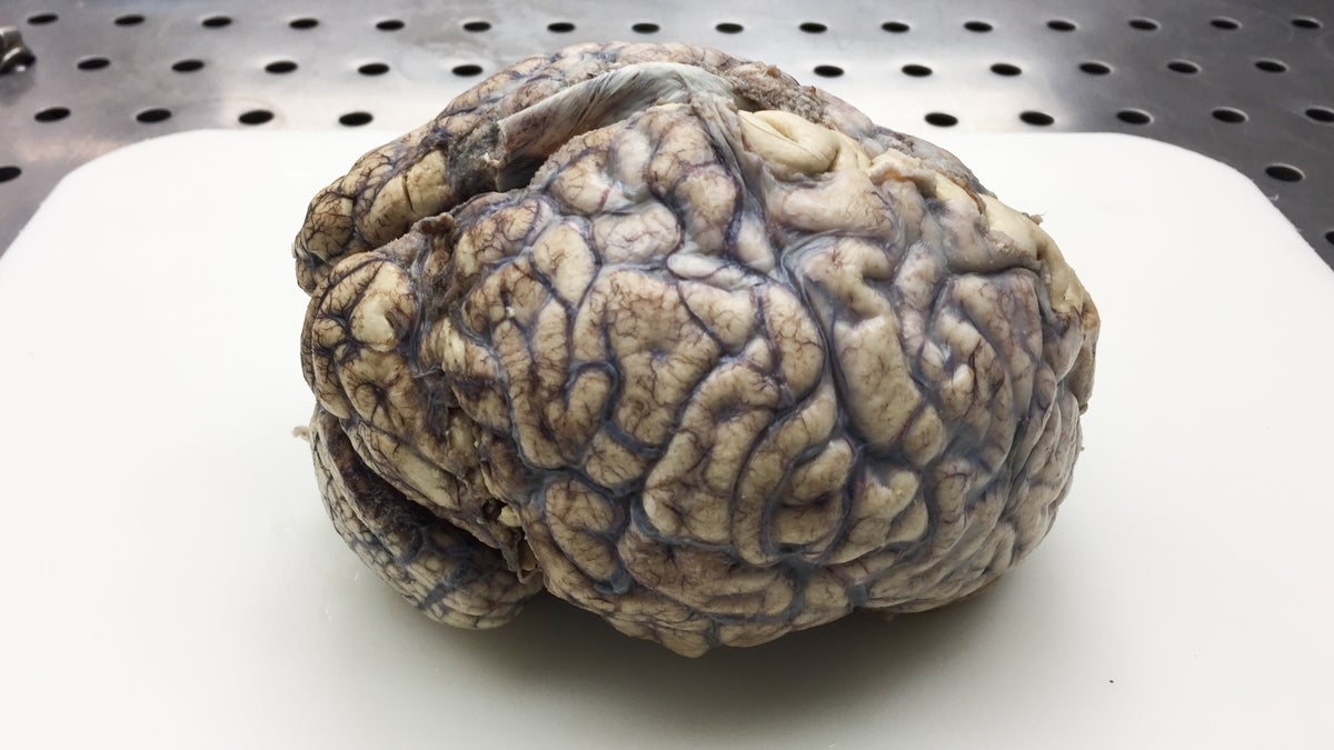
The Brain - WHYY

Neuroanatomy Radiology Reference Article Radiopaedia

Medical Animation Understading orientation of MRI and CT
Diffusion Tensor Imaging 101 Diffusion Imaging

Abnormal Brain MRI Results LIVESTRONG.COM

Study What happens when patients can comparison shop for

The Abnormal Bone Marrow MRI Patterns Radiology Key

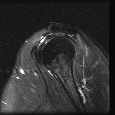
MRI Images - Anatomic Imaging of the Shoulder - MR-TIP.com

Coronal view mri radiograph - Spinal Cord - RR School Of
